Several times over the last couple weeks I have been asked about brainwaves and other measures of the brain. For example, what differentiates a CAT Scan from an EEG, MRI, fMRI, and a PET Scan? And what about those beta, alpha, theta, and delta brain waves? What are they all about? What do these technologies really measure and what can we infer from them?
Before I address these questions, it is important to understand that the brain is an incredibly complicated electrochemical organ composed of an estimated 100 billion neurons (brain cells) interconnected by 100 trillion synapses. Brain activity occurs through a complex interplay of electrical activity within each cell and chemical activity across the synapses (minute gaps between neurons). Once a neuron fires it sends an electrical signal the length of the cell where it releases specific chemicals (called neurotransmitters) into the gap between itself and neighboring neurons. Those neurotransmitters may traverse the gap and attach to neighboring cells’ dendrites (nerve firing receptors), and perhaps trigger a continuation of the signal (via electrical responding) within those adjacent neurons. Every thought we have, every sight we see, everything we feel, taste, and smell occurs through this chain of events.
Obviously, the complexity of this series of events are beyond the scope of this article, but it is important to understand this basic and fundamental fact before one can hope to differentiate the various brain measures. It is also important to understand that there are a number of biological systems that service this neuronal network (e.g., glial cells, veins, and arteries). The glial cells in particular are referred to as “housekeeping” cells protecting, supporting, providing nutrition for, and facilitating communication among the neurons. These cells are the most abundant cells in the brain, but they are not considered to be neurons (MedicineNet, 2004).
At a basic level, brain activity can be measured by the apparent electrical conductivity going on among the neurons. This is what an Electroencephalography (EEG) measures. A series of electrodes are placed on the scalp where they detect and measure this electrical activity. EEGs have been used for years for diagnostic purposes primarily for epilepsy. Formerly, this technique had been used for measuring the impact of strokes and tumors. This function has been replaced by more sophisticated technologies that image the brain (CAT and MRI Scans).
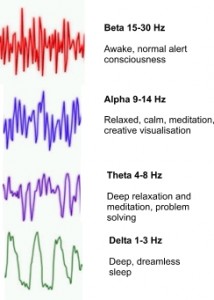
EEGs are relatively inexpensive but valid measures of brain activity. This technology was used in the research I discussed in my last post (The Effect of Low SES on Brain Development), but they can also detect brain death. When someone refers to brain waves, they are referencing the brain activity measured via EEG. Various states of arousal are associated with specific patterns of electrical activity that when measured denote specific wave patters. Those wave patterns are widely known as Beta, Alpha, Theta, and Delta as shown in the image on the right along with the associated arousal states.
These universal brain wave states reflect neuronal activity levels associated with cognitive and bodily activity levels. If you are sleeping yet not dreaming, your brain’s activity level is likely to be represented by high amplitude low frequency Delta waves. At the other extreme, an awake and alert state is likely to be indicated by high frequency Beta waves. These states are not mutually exclusive and any may coexist at any time based on arousal levels.
Much pseudoscience focuses on selling strategies to accomplish “preferred” brain wave states, with little actual data to substantiate claims. Proceed with caution in this realm.
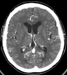
Whereas the output of an EEG is a series of wavy lines, newer technologies actually allow us to image the brain itself or at least proxies of activity. A computerized axial tomography scan, a CAT or CT scan, is an x-ray procedure that combines multiple x-ray images with the aid of a computer to generate cross-sectional views of the internal organs and structures of the body. The image at right is a CAT Scan from Cedars-Sinai.
“Imagine the body as a loaf of bread and you are looking at one end of the loaf. As you remove each slice of bread, you can see the entire surface of that slice from the crust to the center. The body is seen on CT scan slices in a similar fashion from the skin to the central part of the body being examined. When these levels are further “added” together, a three-dimensional picture of an organ or abnormal body structure can be obtained” (MedicineNet.com, 2011).
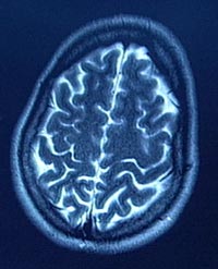
Although CT technology is helpful to look at the structure of the brain and to identify pathologies, it is just a snap shot in time, giving no information about the processes going on within the structure itself. This is also the case for MRI Scans. Non-invasive Magnetic Resonance Imaging (MRI) uses powerful magnets, radio frequency pulses, and a computer to produce very detailed pictures of organs, soft tissues, bone and virtually all other internal body structures without ionizing radiation (x-rays). MRIs provide higher resolution images than x-ray, ultrasound or CAT scans (RadiologyInfo.org).
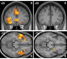
But, if you want to know what is going on in the recesses of the brain you need a functional MRI (fMRI) or a PET scan. The fMRI uses the same technology as an MRI but rather than creating a structural image of the tissue itself, the fMRI looks at blood flow in the brain in order to identify, in real-time, specific locations of brain activity associated with thoughts, external stimuli, or activity. Changes in blood flow captured on a computer, help scientists understand how the brain works. The image above and to the right are fMRI scans showing brain activity in empathy-generating centers of the limbic system in normal individuals (left) and in psychopathic individuals (right) when they are exposed to violent images (Credit: Department of Clinical and Cognitive Neuroscience, University of Heidelberg).

The image on the left shows areas of brain activity associated with being in passionate love (“Graphic Science: Your Brain in Love” in the February 2011 issue of Scientific American. Graphic by James W. Lewis, West Virginia University).
The current state of the art is this fMRI technology because of its superior resolution relative to a PET Scan that formerly comprised the only imaging technology that also indicated brain activity.
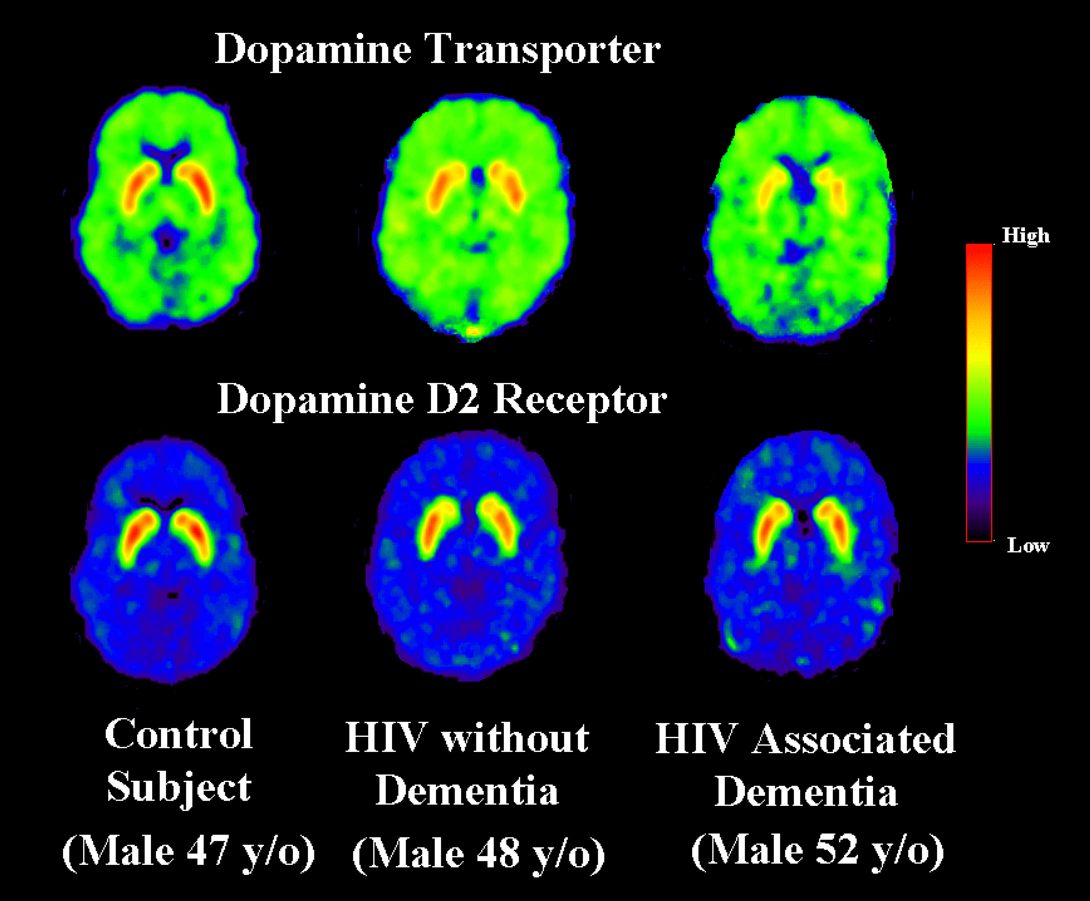
PET Scans or Positron Emission Tomography, is a metabolic imaging tool that is based on molecular biology. PET scan images detail biochemical changes in the body’s tissues, as it traces the body’s metabolic activity. Unlike the newer fMRI technology, PET scans necessitate injections of radioactive material into the body. Since brain activity involves an increase in blood flow, more blood and radioactive material is reflected in the subsequent images. The differences between PET and fMRI scans can be seen by comparing the PET scan image below and the fMRI images above. The PET Scan below was published by scientists at the U.S. Department of Energy’s Brookhaven National Laboratory. They discovered a key mechanism in the brains of people with human immunodeficiency virus (HIV) dementia. The study is the first to document decreases in the neurotransmitter dopamine in those with the condition, and may lead to new, more effective therapies (see HIV Dementia Mechanism Discovered).
This article spans the current brain measuring, imaging, and mapping technologies. Each approach has specific applications and unique advantages and disadvantages. The major advantages of MRI technology include the resolution of its images and the fact that use does not involve x-rays or any radioactive dyes or contrasting agents. However, because MRI machines use 12 to 35 ton magnets, individuals with ferrous metal implants are necessarily excluded from MRI scans for obvious reasons. There are other devices out there with variations on the MRI theme, each serving very unique imaging niches. I won’t go into those here.
An MRI costs on average between $1100 and $2700 (depending on geographical location, facility, and body area imaged), while a CAT Scan costs between $700 and $3000. An fMRI costs up to $2000 per imaging hour. PET scans run between $3000 to $7000. These costs do explain in part why wide-spread use of these imaging technologies are not common in large research projects. They also obviously contribute to the high costs of medical care. But, oh what wonders they offer in our efforts to understand the most complicated thing known to humankind.
References:
Brandt, R. (2007). What can Neuroscience tell us about evil. Technology Review. MIT
Brookhaven National Laboratory. (2004). HIV Dementia Mechanism Discovered. Brookhaven National Laboratory News.
Cedars-Sinai. (2011). CT Brain with or without Contrast.
Fischetti, M. (2011). Passionate Love in the Brain, as Revealed by MRI Scans. Scientific American. “Graphic Science: Your Brain in Love” February 2011 issue.
Howard Hughes Medical Institute. (2008). New Imaging Techniques That Show the Brain at Work: Brain Scans That Spy on the Senses.
MedicineNet.com. (2011). Computerized Axial Tomography.
MedicineNet.com. (2004). Definition of Glial Cell.
RadiologyInfo.org. (2010). MRI of the Head.

Pingback:Tweets that mention Brain Waves and Other Brain Measures » How Do You Think? - A personal exploration of science, skepticism, and how we think. -- Topsy.com
Pingback:What happens in the brain during sensory deprivation? - Quora
Pingback:2011- A Year in Review: How Do You Think? – How Do You Think?
As far as fMRI is concerned even promoters of the technology have conceded that all previous fMRI papers may have to be withdrawn. In a study published 14 October, researchers reanalyzed data from several of their own functional connectivity studies after correcting for head motion and found that this maturation pattern usually disappears once head motion is taken into account.
“It really, really, really sucks. My favorite result of the last five years is an artifact,” says lead investigator Steve Petersen, professor of cognitive neuroscience at Washington University in St. Louis.
It’s unclear how many published results head motion has skewed, and whether this changes the bottom-line conclusions. But many researchers are concerned.
“It’s going to impact some findings with regard to the robustness, but whether it completely wipes out the findings that are out there is another question,” says Damien Fair, assistant professor of behavioral neuroscience and psychiatry at Oregon Health and Science University. “It is going to require folks to reanalyze their data, controlling for these new ways of examining motion.”
Follow the discussion going on at SFARI Autism:
http://sfari.org/news-and-opinion/news/2011/movement-during-brain-scans-may-lead-to-spurious-patterns
Thanks for sharing this information RAJensen. I did look at the link you sent. It seems however that the problem is only pertaining to:
I believe we need be careful not to implicate all fMRI studies. The Functional Connectivity MRI technique employs fMRI technology but in a particular way that maps how brain regions interact. The article references how head movement may lead to spurious results specifically when functional connectivity is assessed in short intervals.
Those interested in more information on developing brain imaging technologies might be interested in HEAD SHOTS in the November/December 2011 Issue of Scientific American Mind By Ann Chin and Sandra Upson. Very interesting stuff – albiet cursory.
Pingback:2012 – A Year in Review: How Do You Think? - How Do You Think?
Pingback:2013 – A Year in Review: How Do You Think? - How Do You Think?